
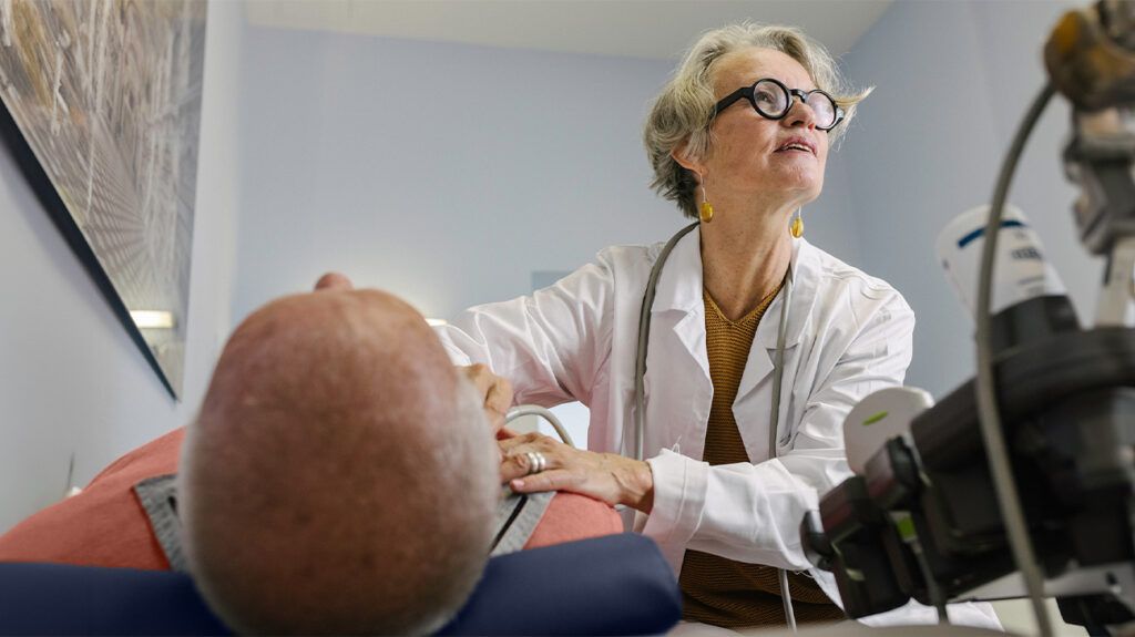
An ultrasound scan is one of the main tests doctors use to diagnose cholecystitis. On a scan, clinicians may be able to spot the presence of gallstones, which can cause this condition.
Cholecystitis is inflammation of the gallbladder. It usually occurs when a gallstone gets stuck at the gallbladder’s opening.
This condition typically causes sharp pain on the upper right-hand side of your abdomen. Without treatment, it can lead to severe complications, so making an accurate diagnosis is important.
In this article, we look at the use of ultrasound imaging in the diagnosis of cholecystitis. We also examine what people can expect if they are due to have this test.
People may receive an ultrasound scan if a doctor thinks that they might have cholecystitis.
Ultrasounds of the gallbladder are the
While ultrasounds are suitable for showing gallstones, they are
Less common causes include injury and severe infection.
Learn more about the causes of cholecystitis.
Before having an ultrasound, a doctor will perform a physical examination to look for other signs of gallbladder inflammation. In particular, they will look for Murphy’s sign to determine whether something is wrong with the gallbladder.
To test for Murphy’s sign, the following will happen:
- The doctor will apply pressure below the patient’s ribs on the right-hand side.
- They will ask the person to breathe in deeply.
- The gallbladder will move close to where the doctor has placed their hand.
- If the gallbladder is inflamed, this process will be painful, and the person will be unable to inhale further.
Doctors may also order blood tests. If the gallbladder is inflamed, the person will likely have a
If someone needs to have an ultrasound, they may have this at the doctor’s office or the hospital. They will not need to do any preparation beforehand.
Sonographers, who are healthcare professionals who use ultrasound imaging, carry out ultrasound scans by placing a wand-like device called a transducer on a person’s skin. They may apply gel to the skin to allow for smooth movement and continuous contact.
The transducer emits sound waves through the body, and an image forms as the sound bounces back off denser surfaces such as gallstones.
Clinicians can also test for Murphy’s sign during the ultrasound.
An ultrasound scan will likely take less than 45 minutes.
Learn more about how ultrasound scans work.
An ultrasound can spot many factors relating to cholecystitis.
- gallstones
- enlargement of the gallbladder
- thickening of the gallbladder wall
- a buildup of fluid around the gallbladder
The presence of these signs can help diagnose cholecystitis.
Ultrasounds are
If these signs of cholecystitis are not present, the symptoms a person is experiencing will likely have a different cause. Conditions with similar symptoms include gastritis and appendicitis.
If a doctor is able to diagnose cholecystitis after the ultrasound, they will decide on the best course of treatment.
In many cases, they will recommend gallbladder removal surgery due to the risk of recurrence. This can be open surgery or keyhole surgery.
If surgery would pose risks for the person and the cholecystitis is a particularly mild case,
If the doctor is unable to make a
Bile is a digestive fluid that the gallbladder stores.
Learn more about gallbladder removal surgery.
What are the ultrasound findings for cholecystitis?
If a person has cholecystitis, an ultrasound will often show the presence of gallstones in the opening of the gallbladder. It may also show if the gallbladder has swollen or if there is a buildup of fluid around it.
What is the best imaging test for cholecystitis?
Clinicians consider ultrasound to be the best imaging test for cholecystitis due to its speed, accessibility, and the fact that it does not use radiation.
Will an inflamed gallbladder show up on an ultrasound?
Yes, in some cases, sonographers will be able to detect gallbladder inflammation on an ultrasound.
Ultrasounds are an important test that clinicians use to diagnose cholecystitis. They can detect the presence of gallstones in the opening of the gallbladder, which is the cause of most cases.
Doctors prefer to use ultrasounds over other forms of imaging because it is quicker and easier than other forms, such as CT scans.
Doctors may also use blood tests and physical examinations to check for Murphy’s sign alongside ultrasounds.