
Doctors may use a range of tests to diagnose myasthenia gravis (MG), including blood tests, nerve conduction tests, and imaging tests.
MG is a chronic autoimmune disease that affects the voluntary muscles, which are the muscles a person can control. Voluntary muscles are involved in various functions, including movement, swallowing, and breathing.
In MG, immune proteins called antibodies mistakenly block or destroy receptors for a neurotransmitter called acetylcholine.
Among other functions, acetylcholine initiates the transmission of signals responsible for movement. As MG affects acetylcholine receptors, this condition impairs signaling between the nerves and muscles. Ultimately, this prevents the muscles from contracting properly.
A neurological exam involves a series of tests to evaluate a person’s sensory and motor neuron responses. This involves assessing the following:
- reflexes
- muscle tone
- muscle strength
- gait
- posture
- coordination
- balance
- senses of touch
- sense of sight
- mental skills
Issues such as muscle weakness or impaired eye movement may warrant further tests for MG.
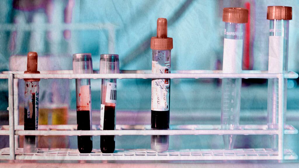
Myasthenia gravis can cause elevated levels of certain antibodies in the blood.
According to Conquer Myasthenia Gravis, around 80% to 85% of individuals with MG test positive for acetylcholine receptor (AChR) antibodies.
Another 5% to 10% test positive for anti-muscle-specific serum kinase (anti-MuSK) antibodies. The MuSK protein helps organize acetylcholine on the surface of muscle cells. Consequently, anti-MuSK antibodies interfere with typical muscle function.
While blood tests can help diagnose MG, it is worth noting that some individuals with this condition have typical levels of antibodies in their blood. This is often the case with MG that is restricted to the eyes.
The ice test is important for diagnosing ocular MG. It uses the principle that cooling the muscles can temporarily reduce muscle weakness in MG.
To perform the ice test, the doctor first assesses the amount of eyelid drooping (ptosis) by measuring the space between the upper and lower eyelid.
They then apply ice packs to one or both eyes for up to 2 minutes.
After removing the ice packs, the doctor rechecks the amount of ptosis in the affected eye. A change of more than 2 millimeters in eyelid opening indicates ocular MG.
Nerve conduction tests measure the speed with which nerves transmit electrical impulses to the muscles or sensory nerves.
To perform a nerve conduction study, a clinician will place two types of electrodes onto specific areas of the skin — stimulating electrodes and recording electrodes.
Stimulating electrodes deliver a mild electrical current through a nerve, and recording electrodes record the muscle’s response to the electrical stimulation.
The clinician then records how long it takes for the muscle to respond to the nerve signal. The medical term for this response is the conduction velocity.
In MG, the muscles take longer to respond to repeated stimulation.
According to a 2021 review, single fiber EMG is the most sensitive test for diagnosing MG.
As the United Kingdom’s National Health Service (NHS) explains, single fiber EMG is an extremely sensitive test for measuring the function of the neuromuscular junction, which is the connection between a muscle and a nerve.
In this test, a clinician inserts a very fine needle into an individual muscle fiber and then asks the person to contract or activate the muscle.
Recording equipment then sends the data to a computer that analyses the electrical signals.
Individuals with MG or other neuromuscular disorders may show abnormal neuromuscular transmission.
Imaging tests, such as CT or MRI scans, can help identify issues with the thymus gland, a small gland in the upper chest that makes immune cells called lymphocytes.
According to the
The thymus gland grows gradually up to puberty and then decreases in size until eventually replaces it. In many people with MG, the thymus gland remains large into and throughout adulthood.
These individuals may develop clusters of immune cells on the thymus gland and thymus tumors called thymomas. The thymus gland may send faulty instructions to developing immune cells, causing them to produce AChR antibodies.
The edrophonium test involves administering a short-acting intravenous (IV) drug called edrophonium chloride (EC). This drug blocks an enzyme that breaks down acetylcholine.
As such, an improvement in muscle weakness following the administration of EC indicates possible MG.
However, according to the NHS, a healthcare professional may only recommend this test if it is unclear what is causing a person’s symptoms. This is because there is a risk of severe side effects, such as breathing problems and slow heartbeat.
Due to the risk of side effects, this test takes place in the hospital where treatment for the side effects is easily available.
A person does not need to make any special preparations for the following tests:
- neurological tests
- the ice pack test
- blood antibody tests
Certain preparations may be necessary for the other tests, such as fasting or avoiding certain foods and beverages.
A person’s doctor will offer specific advice on how to prepare for each test, if necessary.
Neurological tests and ice pack tests are MG tests that do not pose any risk to a person’s health.
Tests that pose very little risk include:
- blood tests, although a person may experience slight pain or temporary bruising at the site of needle insertion
- nerve conductions studies, which may cause slight tingling sensations
- an EMG test, which may cause mild pain or cramping
- an MRI scan, which is painless but may cause feelings of claustrophobia in susceptible individuals
An endrophonium test may cause an allergic reaction, though this is not common. Anyone who experiences symptoms of an allergic reaction during this test should notify the doctor or nurse immediately. Symptoms that may indicate a severe allergic reaction can include:
- tightness of the throat
- difficulty breathing
- nausea or vomiting
- dizziness
- fainting
- rapid heart rate
- a feeling of doom
Myasthenia gravis (MG) is a chronic neurological and autoimmune condition that affects voluntary muscle function. In MG, the immune system mistakenly attacks acetylcholine receptors, impairing nerve signaling and muscle function. People with MG may experience symptoms such as muscle weakness or speech, swallowing, or breathing difficulties.
Various tests can help doctors diagnose MG. The most sensitive of these is single fiber electromyography (EMG), which can detect even mild cases of the disease. Other tests include a neurological examination, blood antibody tests, and imaging tests.
Most diagnostic tests for MG do not require any special preparation. However, some tests may require a person to fast or avoid consuming certain foods or beverages beforehand. A person’s doctor will offer specific guidance on preparing for a particular test and outline any potential risks involved.