
A macular hole may progress through four stages, each with different features. An early stage hole may resolve without treatment, while a later stage hole typically requires surgery.
The macula is a small, light-sensitive area in the back of the eye that enables clear and detailed central vision.
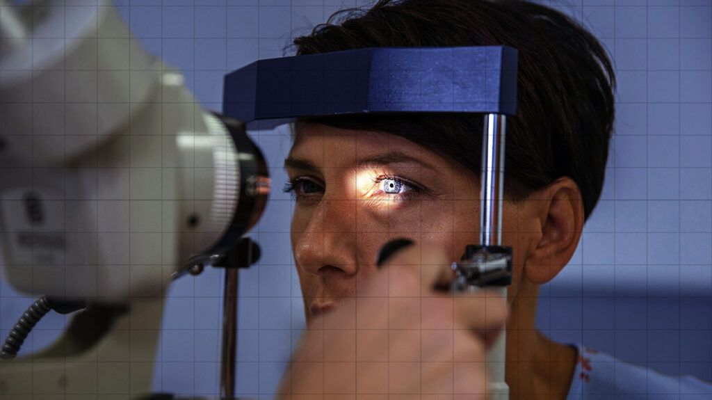
A macular hole is a circular opening in the macula that causes objects in central vision to appear blurry, wavy, or distorted.
A stage 1 macular hole represents the earliest stage of macular disease.
Stage 1A
In stage 1A, the eye’s foveola separates from the center of the macula. The foveola is the part of the macula that contains the
During this stage, tiny yellow protein deposits called drusen may develop beneath the macula. These are visible on color photographs of the retina.
Stage 1B
In a stage 1B hole, the foveola separates from the center of the macula and a yellow ring surrounds the detachment.
However, doctors may recommend surgery for a stage 1 macular hole that significantly and persistently affects how clearly a person can see.
A stage 2 macular hole involves a full-thickness break in the fovea that measures under
During this stage, doctors may recommend a type of eye surgery called vitrectomy, which
The bubble acts as a temporary bandage that holds the edges of the macular hole together during healing.
As the American Academy of Ophthalmology explains, the eye eventually replaces the solution or bubble with a natural fluid called aqueous humor.
A stage 3 macular hole involves a full-thickness break in the fovea measuring more than
During this stage, there may also be early signs of posterior vitreous detachment (PVD), in which the vitreous gel that fills the eye separates from the retina.
Treatment for a stage 3 macular hole is the same as for a stage 2 hole.
A stage 4 macular hole includes all the features of stage 3 but also involves complete PVD, as indicated by the development of a Weiss ring.
According to the American Society of Retina Specialists (ASRS), a Weiss ring is a type of circular or oval-shaped floater that can obscure vision.
The treatment for a stage 4 macular hole is the same as for stage 2 and 3 macular holes.
Learn more about eye floaters.
According to the
At first, a person may not experience any symptoms, especially if they have good vision in the other eye.
When early stage symptoms occur, a person may notice that their central vision becomes blurry or distorted. In particular, lines or straight objects may appear bent, wavy, or as though they are missing a piece in the center.
This can interfere with the following activities:
- reading or writing
- watching television
- driving
As the macular hole progresses, a person may notice a dark or blind spot in their central vision.
When reading, a person may see lines of text but be unable to differentiate words in the center of each line or letters within words. People may also find facial recognition challenging as they become less able to distinguish facial features.
Doctors may recommend a “watch and wait” approach to see if an early stage macular hole progresses or heals without treatment.
Vitrectomy is the standard treatment for a macular hole that does not resolve spontaneously. According to the ASRS, over 90% of people who receive this treatment regain some or most of their vision.
Nonetheless, surgical complications are possible and may include:
Around 10% of people who undergo vitrectomy find that their macular hole persists or reopens. These individuals may require follow-up surgery.
Another potential treatment option is a medication called ocriplasmin (Jetrea), which doctors inject directly into the vitreous. This helps stop the vitreous from pulling on the macula. Rarely, this treatment can cause severe side effects, such as:
- noticeable vision loss
- macular hole enlargement
- retinal detachment
According to the United Kingdom’s National Health Service, people should contact a healthcare professional if they experience any of the following potential symptoms of a macular hole:
- blurred vision
- distorted vision
- a dark spot in the center of the visual field
Without a diagnosis and appropriate treatment, a macular hole may worsen, causing permanent vision impairment. Early treatment can help restore vision, though a watch and wait approach is more appropriate in some cases.
A person should also contact a healthcare professional if their vision problems persist or worsen following treatment for a macular hole. They may require follow-up treatment to correct any underlying issues.
How quickly does a macular hole progress?
The rate of macular hole progression is highly variable. However, early treatment is more effective.
A
- Small: 1.67 microns per day
- Medium: 0.61 microns per day
- Large: 0.44 microns per day
When is it too late to treat a macular hole?
According to the
How urgent is macular hole surgery?
The urgency of macular hole surgery may differ from person to person.
For a stage 1 macular hole, doctors may recommend a watch and wait approach. Around
Later stage macular holes require more urgent treatment to help prevent permanent vision loss.
Eye health resources
Visit our dedicated hub for more research-backed information and in-depth resources on eye health.
A macular hole is a circular opening in the eye’s macula that causes central vision to become blurred or distorted. Macular holes may progress through four distinct stages, gradually causing a progressive loss of central vision.
Stage 1 macular holes may resolve without treatment. However, later stage macular holes require treatment, such as vitrectomy or injectable medications, to help prevent permanent vision loss.
Anyone with symptoms of a macular hole needs to consult a doctor or ophthalmologist for a diagnosis and appropriate treatment. Early treatment can lead to a better outcome in terms of retaining vision.