
Choroid plexus tumors are growths in the area of the brain that produces cerebrospinal fluid (CSF). They may be cancerous or noncancerous.
Choroid plexus tumors are rare. They develop in the choroid plexus, a network of cells lining the brain’s ventricles. Tumors in this area can disrupt the flow and production of CSF, leading to various neurological symptoms.
Although these tumors can affect individuals of all ages, the benign type most commonly develops during childhood. Early detection and appropriate treatment are crucial for improving outcomes and managing the symptoms of these tumors.
This article examines choroid plexus tumors, their causes, symptoms, and treatments.
Choroid plexus tumors can be either benign or malignant. In a 2024 analysis of over 600 people with these tumors from the years 2000 to 2019, over 70% of the tumors were benign, or noncancerous.
However, some choroid plexus tumors are cancerous. When they are, the tumors can be fast-growing and more difficult to treat.
Doctors grade choroid plexus tumors based on their appearance under a microscope and their potential to grow and spread. The grades are
- Grade 1: These tumors are also known as choroid plexus papillomas (CPPs). They are not cancerous and grow slowly.
- Grade 2: These are atypical CPPs. They are also noncancerous, but they are more likely to come back in the same location after removal than grade 1 tumors.
- Grade 3: These tumors are known as choroid plexus carcinomas. They are cancerous and have a higher tendency to proliferate and spread to other parts of the central nervous system (CNS).
Choroid plexus tumors can increase intracranial pressure as they disrupt the flow of CSF, leading to a condition known as hydrocephalus.
Hydrocephalus may cause:
- headaches
- irritability
- nausea or vomiting
- drowsiness or lethargy
Other symptoms of choroid plexus tumors
- meningitis-like symptoms, such as a stiff neck, light sensitivity, and a headache
- seizures
- weakness or difficulty moving one side of the body
- facial drooping, weakness, or spasms
- changes in vision
People with any of the above symptoms should seek urgent medical help, as these symptoms can be a sign of a serious condition, such as bleeding in the brain, meningitis, or stroke.
The exact cause of choroid plexus tumors is unknown, but research has identified several factors that may increase the risk of developing them, including:
- Genetic predisposition: There is an association between certain genetic conditions and a higher risk of developing choroid plexus tumors. They include Li-Fraumeni syndrome, Aicardi syndrome, Gorlin syndrome, and familial adenomatous polyposis.
- Age: CPPs are more prevalent in children, with
70% of diagnoses occurring in children under the age of 2. The reason for this is not fully understood, but it may be related to developmental factors in the brain during early childhood. - Environmental factors: While the primary causes are mainly genetic,
researchers are investigating whether environmental exposures, such as radiation exposure, might contribute to the development of these tumors.
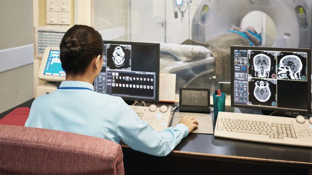
Diagnosing choroid plexus tumors involves a combination of neurological examinations, imaging tests, and sometimes, biopsy procedures.
Doctors begin with a thorough neurological examination to assess the individual’s symptoms and overall neurological function. This includes checking reflexes, muscle strength, coordination, balance, and cognitive abilities.
Next, a doctor may recommend imaging studies, such as:
- MRI scan: This is the preferred imaging method for choroid plexus tumors as it provides more detailed images of the brain’s structures. Additionally, an MRI often shows flow voids within the tumor, indicating active blood flow, a characteristic feature of CPPs.
- CT imaging: For CPPs, CT imaging may show a well-defined, lumpy mass that can appear similar to or more dense than usual brain tissue.
For some people, doctors may perform a biopsy to obtain a tissue sample from the tumor. This involves removing a small portion of the tumor for examination under a microscope. The biopsy helps determine the type and grade of the tumor, providing crucial information for planning treatment.
Treatment for choroid plexus tumors depends on the type, grade, and location of the tumor. The options may include:
- Surgery: When possible, the main treatment for choroid plexus tumors is surgical removal. Surgery aims to remove as much of the tumor as possible without causing further symptoms.
- Radiation therapy: This involves using radiation to kill cancer cells. Doctors may use it after surgery to remove cells that surgery did not.
- Chemotherapy: These potent drugs target cancerous cells and prevent recurrence. Similarly to radiation, doctors can also use it after surgery.
People may also be able to take part in clinical trials for other types of treatment, such as targeted therapies or immunotherapy.
The prognosis for choroid plexus tumors varies based on the tumor’s type and grade.
A large 2024 analysis of people with choroid plexus tumors from the year 2000 to 2019 found overall 5-year survival rates of:
- 90% for grade 1 tumors
- 79% for grade 2 tumors
- 61% for grade 3 tumors
In the data, there was no association between surgical removal and better outcomes for grade 1 tumors, but there was for grade 3.
In those with grade 3 tumors, both complete surgical removal and partial removal with additional cancer therapies had links with higher survival rates.
It is important to note these statistics do not predict survival in an individual’s case. Additionally, this data comes from a span of nearly 20 years. As cancer treatments are always improving, these figures may not accurately represent survival rates today.
Choroid plexus tumors are
Symptoms may include headaches, nausea, vomiting, balance or coordination problems, and seizures. Treatment typically includes surgical removal, radiation therapy, or chemotherapy, depending on whether the tumor is malignant.
Low grade choroid plexus tumors usually have a good prognosis, but grade 3 malignant tumors have a more variable outlook. Early intervention and a comprehensive, multidisciplinary approach are crucial for managing symptoms and improving outcomes.