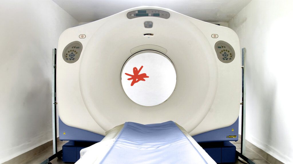
MRI scans create detailed images of soft tissues and tumors, making them highly effective for detecting cancer in various parts of the body. However, they have limitations and may not detect all types of cancer.
Magnetic resonance imaging (MRI) is a powerful and noninvasive diagnostic tool that enables doctors to see inside the body. It uses magnetic fields and radio waves to generate detailed images of organs, tissues, and other structures, including tumors.
This article explores the capabilities and limitations of MRI in cancer detection, comparing it with other imaging tests, and discusses what happens during an MRI procedure.
Cancer resources
To discover more evidence-based information and resources for cancer, visit our dedicated hub.

An MRI scan can assist doctors in detecting cancer in various parts of the body due to its ability to distinguish between different types of tissues. For example, these scans can detect:
- brain tumors
- primary bone tumors
- soft tissue sarcomas
- spinal cord tumors
- prostate cancer
- bladder cancer
- ovarian cancer
MRIs can also check for signs that cancer has spread from its original location. Analyzing MRI results helps doctors plan cancer treatments, such as surgery or radiation therapy.
Despite its versatility, MRI has limitations. For example, it is
This is because air and bowel motion in these areas can interfere with image clarity. That said, technological advancements and breath-holding techniques
MRI may have less success in accurately detecting tiny tumors than other imaging techniques.
Learn about screening for lung cancer using CT scans.
Full-body scans can detect cancer, but doctors typically reserve them for specific medical situations.
Whole-body MRI (WB-MRI) is a scanning method that does not use harmful radiation and can cover the entire body in
WB-MRI is often more effective than bone scans and CT scans for finding and assessing lesions, checking how well treatments are working, and screening people at high risk. Doctors may use additional scans to focus on specific areas if necessary.
Because of its effectiveness, doctors can perform WB-MRI to manage:
- multiple myeloma
- prostate cancer
- melanoma
- genetic cancer risks
Doctors are using it increasingly to identify:
However, the
WB-MRIs should not replace routine screening procedures or targeted imaging tests.
Learn whether a biopsy or MRI is better for prostate cancer.
An MRI requires very little preparation. Before the procedure, a health professional will check there is no medical reason that a person cannot undergo the scan. They may do this by asking them to fill out a questionnaire.
Then, they may ask the person to change into a hospital gown and remove any metal from their body, such as jewelry. As an MRI involves strong magnets, it is critical there are no metal objects in the scanner.
Once ready, the scan will involve the following steps:
- The person will lie on a motorized table. A technician may place blocks around the part of the body they are focusing on to ensure it stays still. They may also give a person a button they can press if they need help and may provide earplugs to reduce the noise of the scanner.
- Next, the table slides into a large tube-shaped MRI machine, and the scan will begin.
- The machine generates a strong magnetic field around the area of interest and sends radio waves through the body. This process takes some time and can be loud.
- The machine’s sensors detect the energy emitted by hydrogen atoms in the body’s cells. These signals are processed to create detailed images of the internal structures.
The procedure is noninvasive and typically lasts
Sometimes, medical professionals inject a contrast dye through the vein to enhance the visibility of certain tissues or blood vessels.
It is important to remain still during the scan to ensure high-quality images. If a person feels uncomfortable during the procedure, they can speak to the MRI technician or press the button in their hand to stop it.
Learn about the side effects of MRI contrast.
MRI and CT scans are valuable tools for detecting cancer, each having distinct advantages and limitations.
MRI
MRI scans can produce
An MRI is ideal for scanning the brain, spinal cord, and musculoskeletal system. Additionally, it does not use ionizing radiation, making it a safer option for multiple uses.
However, it can be
CT
CT scans use X-rays to create detailed cross-sectional images of the body. They are
CT scans are also faster than MRIs, and doctors often prefer them in emergency settings. However, a CT scan involves exposure to ionizing radiation, which can accumulate if a person has many scans over time.
The choice between MRI and CT depends on various factors, such as the area of the body a doctor is examining and a person’s specific needs. Doctors will consider these factors when deciding whether to use an MRI or CT scan.
Learn about the differences between MRI and CT scans.
While MRIs are highly effective in detecting and characterizing tumors, they
However, MRIs can provide clues as to whether the tumor might be cancerous based on certain factors, such as its:
- size
- shape
- location
- interaction with surrounding tissues
Benign tumors
A firm diagnosis typically requires a biopsy, where a doctor examines a tissue sample from the tumor under a microscope. MRI findings can guide the biopsy by identifying the areas of a tumor to sample.
Learn more about the different types of tumors.
If an MRI detects a mass that could be cancer, the next steps usually involve further diagnostic testing and a consultation with a specialist. This may include additional imaging tests, such as CT or PET scans, or a biopsy to gather more information about the tumor.
A multidisciplinary team of healthcare providers will use these findings to develop a tailored treatment plan. Treatment options may include:
- surgery
- radiation therapy
- chemotherapy
- targeted therapy
- a combination of these approaches
It is important to note that the outlook for cancer varies widely depending on the type and stage. Some types are highly treatable and are rarely fatal with appropriate care. Others are more difficult to treat, particularly if they are more advanced.
People can speak with their doctor about what their MRI results mean, what their diagnosis might be, and the available treatments.
MRI is a powerful imaging tool that can help detect cancer in various parts of the body. It is particularly effective for scanning soft tissues in the brain, spinal cord, and musculoskeletal system.
However, its effectiveness varies, and it can be less effective for detecting lung and gastrointestinal cancers. It also
The choice between MRI and CT scans depends on an individual’s needs, with each imaging type offering unique strengths. While full-body MRI scans are available, doctors generally do not recommend them for people without any symptoms.
If a doctor detects cancer, further tests and consultations will guide the treatment plan. Individuals should discuss their specific medical needs with their healthcare team.
